3.1 Neurons, Neurotransmitters, and Hormones
Learning Objectives
- Describe the structure and functions of the neuron.
- Draw a diagram of the pathways of communication within and between neurons.
- List three of the major neurotransmitters, and describe their functions.
The nervous system is composed of more than 100 billion cells known as neurons. A neuron is a cell in the nervous system whose function it is to receive and transmit information (see Figure 3.1). Neurons are made up of three major parts: a cell body, or soma, which contains the nucleus of the cell and keeps the cell alive; a branching treelike fibre known as the dendrite, which collects information from other cells and sends the information to the soma; and a long, segmented fibre known as the axon, which transmits information away from the cell body toward other neurons or to the muscles and glands.
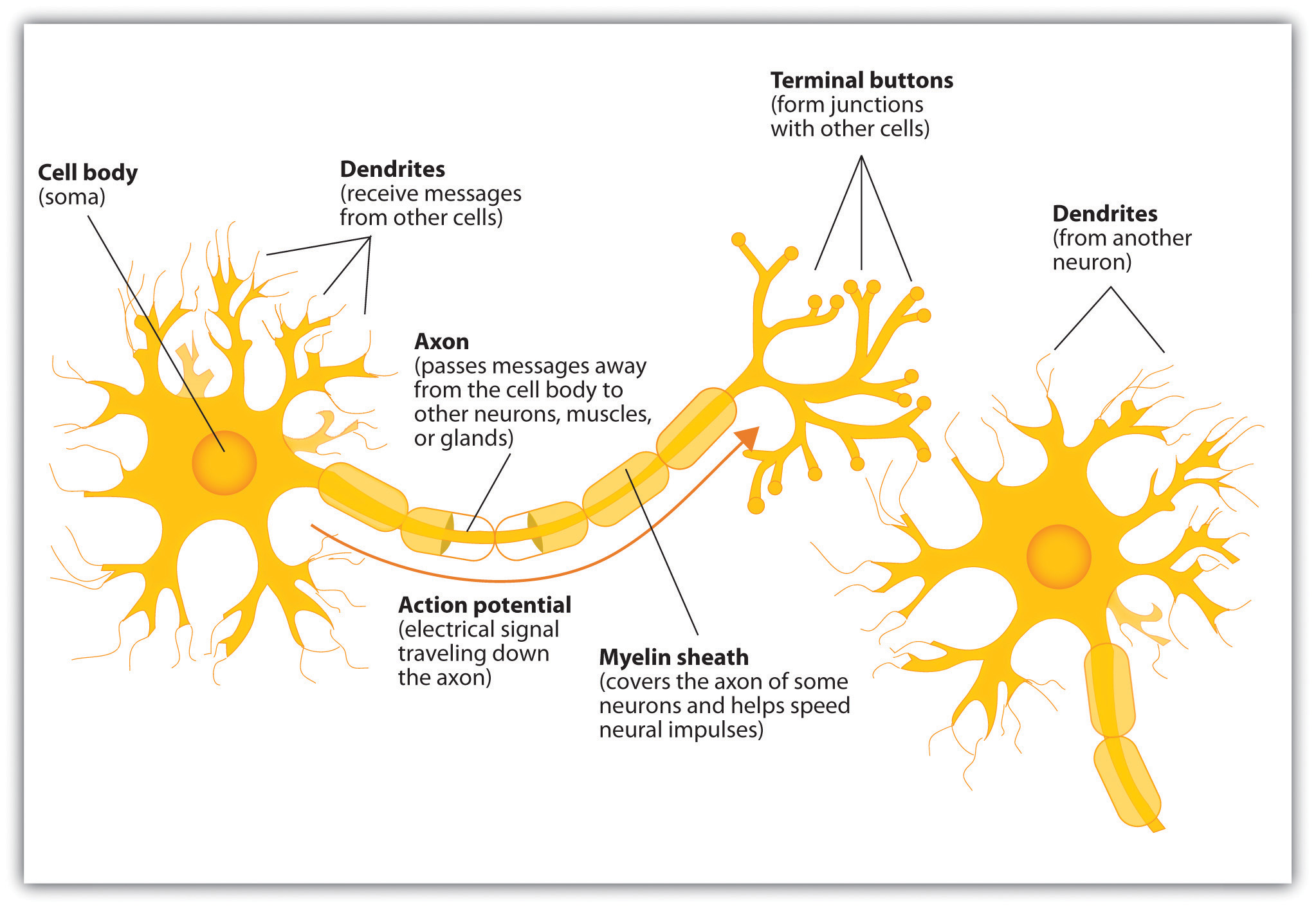
Some neurons have hundreds or even thousands of dendrites, and these dendrites may themselves be branched to allow the cell to receive information from thousands of other cells. The axons are also specialized, and some, such as those that send messages from the spinal cord to the muscles in the hands or feet, may be very long — even up to a metre in length. To improve the speed of their communication and to keep their electrical charges from shorting out with other neurons, axons are often surrounded by a myelin sheath. The myelin sheath is a layer of fatty tissue surrounding the axon of a neuron that both acts as an insulator and allows faster transmission of the electrical signal. The myelin sheath is segmented, which allows for more efficient transmission of information down the axon. The axon is segmented by a series of breaks between the sausage-like segments of the myelin sheath (see Figure 3.2). Each of these gaps is a node of Ranvier. Axons branch out toward their ends, and at the tip of each branch is a terminal button.

The following YouTube link provides an excellent explanation of the parts of a neuron:
- Video: Anatomy of a Neuron | Human Anatomy and Physiology | Health & Medicine | Khan Academy (Khan Academy, 2010)
Neurons communicate using electricity and chemicals
The nervous system operates using an electrochemical process. An electrical charge moves through the neuron itself, and chemicals are used to transmit information between neurons. Within the neuron, when a signal is received by the dendrites, it is transmitted to the soma in the form of an electrical signal, and, if the signal is strong enough, it may then be passed on to the axon and then to the terminal buttons. If the signal reaches the terminal buttons, they are signaled to emit chemicals known as neurotransmitters, which communicate with other neurons across the spaces between the cells, known as synapses. Thus, communication between neurons is both electrical and chemical.
The electrical signal moves through the neuron as a result of changes in the electrical charge of the axon. When an axon is in a state of rest, the electrical charge is said to be negative because the interior of the neuron contains a greater number of negatively charged ions than the area outside of it. This state is called the resting potential. The resting potential is generally about -70 millivolts (mV).
When a neuron receives stimulation via its dendrites, there is a change in the electrical charge of the receiving neuron. If this electrical signal is strong enough that it passes a certain level, or threshold, of approximately -55 mV, the membrane of the segment of the axon closest to the cell body opens its gates, allowing positively charged sodium ions to enter that were previously kept out. In other words, the previous resting potential of -70 mV has changed and become more positive, to approximately -55 mV. With the firing threshold reached, a wave of electrical activity will rapidly travel down the axon. This change in electrical charge that occurs in a neuron when a nerve impulse is transmitted is known as the action potential. Once the action potential occurs, the number of positive ions exceeds the number of negative ions in this segment, and the segment temporarily becomes positively charged.
The electrical charge moves down the axon from segment to segment in a set of small jumps, moving from node to node. When the action potential occurs in the first segment of the axon, it quickly creates a similar change in the next segment, which then stimulates the next segment, and so forth as the positive electrical impulse continues all the way down to the end of the axon. As each new segment becomes positive, the membrane in the prior segment closes up again, and the segment returns to its resting potential. In this way, the action potential is transmitted along the axon toward the terminal buttons. The entire response along the length of the axon is very fast; it can happen up to 1,000 times each second.
An important aspect of the action potential is that it operates in an all-or-nothing manner. What this means is that the neuron either fires completely, such that the action potential moves all the way down the axon, or it does not fire at all. Thus, neurons can provide more energy to other neurons down the line by firing quickly, not by firing more powerfully. Furthermore, the neuron is prevented from repeated firing by the presence of a refractory period, which is a brief time after the firing of the axon in which the axon cannot fire again because the neuron has not yet returned to its resting potential.
The following YouTube link explains the details of the action potential:
- Video: The Nervous System, Part 2 – Action! Potential!: Crash Course A&P #9 (CrashCourse, 2015)
Neurotransmitters
Communication between neurons is chemical. Neurons are separated by synapses, the small gap between neurons across which nerve impulses are transmitted. The synapse is where the terminal buttons at the end of the axon of one neuron nearly, but do not, touch the dendrites of another. Synapses provide a remarkable function because they allow each axon to communicate with many dendrites in neighbouring cells. Because a neuron may have synaptic connections with thousands of other neurons, the communication links among the neurons in the nervous system allow for a highly sophisticated communication system.
When the electrical impulse from the action potential reaches the end of the axon, it signals the terminal buttons to release neurotransmitters into the synapse. A neurotransmitter is a chemical that relays signals across the synapses between neurons. Neurotransmitters travel across the synaptic space between the terminal button of one neuron and the dendrites of other neurons where they bind to the dendrites in the neighbouring neurons (see Figure 3.3). Furthermore, different terminal buttons release different neurotransmitters, and different dendrites are particularly sensitive to different neurotransmitters. The dendrites will admit the neurotransmitters only if they are the right shape to fit in the receptor sites on the receiving neuron. For this reason, the receptor sites and neurotransmitters are often compared to a lock and key.
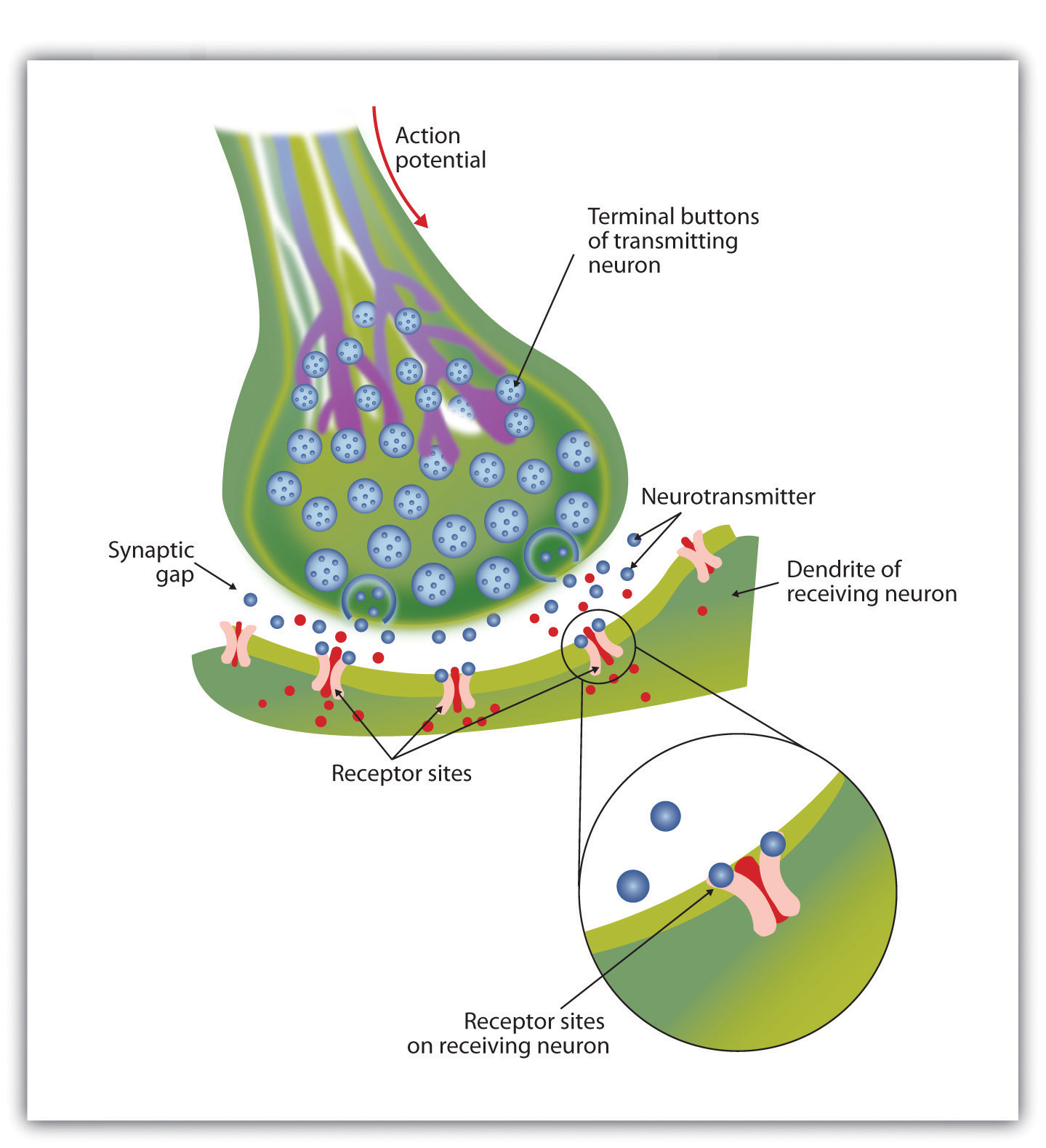
When neurotransmitters are accepted by the receptors on the receiving neurons, their effect may be either excitatory (i.e., they make the cell more likely to fire) or inhibitory (i.e., they make the cell less likely to fire). Furthermore, if the receiving neuron is able to accept more than one neurotransmitter, it will be influenced by the excitatory and inhibitory processes of each. If the excitatory effects of the neurotransmitters are greater than the inhibitory influences of the neurotransmitters, the neuron moves closer to its firing threshold; if it reaches the threshold, the action potential and the process of transferring information through the neuron begins.
After an action potential occurs, neurotransmitter molecules must be removed from the synapse in order for the next potential stimulation of the neuron to happen. This process occurs, in part, through the breaking down of the neurotransmitters by enzymes and, in part, through reuptake, which is a process in which neurotransmitters that are in the synapse are reabsorbed into the transmitting terminal buttons, ready to be released again after the neuron fires. Some medications inhibit the reuptake of neurotransmitters, allowing the neurotransmitter to remain in the synapse for longer and increasing its effectiveness.
More than 100 chemical substances produced in the body have been identified as neurotransmitters, and these substances have a wide and profound effect on emotion, cognition, and behaviour. Neurotransmitters regulate our appetite, our memory, our emotions, as well as our muscle action and movement. As you can see in the table below, some neurotransmitters are also associated with psychological and physical diseases.
| Neurotransmitter | Description and Function | Notes |
|---|---|---|
| Acetylcholine (ACh) | Stimulates muscle contractions; also used in the brain to regulate memory, sleeping, and dreaming. | Alzheimer’s disease is associated with an undersupply of acetylcholine. Nicotine is an agonist that acts like acetylcholine. |
| Dopamine | Involved in movement, motivation, and emotion; produces feelings of pleasure when released by the brain’s reward system; also involved in learning. | Schizophrenia is linked to increases in dopamine, whereas Parkinson’s disease is linked to reductions in dopamine, so dopamine agonists may be used to treat it. |
| Endorphins | Released in response to behaviours such as vigorous exercise, orgasm, and eating spicy foods. | Endorphins are natural pain relievers. They are related to the compounds found in drugs such as opium, morphine, and heroin. The release of endorphins creates a sense of euphoria (e.g., the runner’s high) that is experienced after intense physical exertion. |
| Gamma-aminobutyric acid (GABA) | The major inhibitory neurotransmitter in the brain; lowers arousal; involved in sleep. | A lack of GABA can lead to involuntary motor actions, including tremors and seizures. Alcohol stimulates the release of GABA, which inhibits the nervous system and makes us feel drunk. Low levels of GABA can produce anxiety, and GABA agonists (e.g., tranquilizers) are used to reduce anxiety. |
| Glutamate | The most common neurotransmitter; an excitatory neurotransmitter released in more than 90% of the brain’s synapses. | Excess glutamate can cause overstimulation, migraines, and seizures. |
| Serotonin | Involved in many functions, including mood, appetite, sleep, and aggression. | Low levels of serotonin are associated with depression. Some drugs designed to treat depression, known as selective serotonin reuptake inhibitors (SSRIs), serve to prevent their reuptake. |
Drugs that we might ingest — either for medical reasons or recreationally — can act like neurotransmitters to influence our thoughts, feelings, and behaviour. An agonist is a drug that has chemical properties similar to a particular neurotransmitter, which allows it to mimic the effects of the neurotransmitter. When an agonist is ingested, it binds to the receptor sites in the dendrites to excite the neuron, acting as if more of the neurotransmitter had been present. As an example, cocaine is an agonist for the neurotransmitter dopamine. Because dopamine produces feelings of pleasure when it is released by neurons, cocaine creates similar feelings when it is ingested. An antagonist is a drug that reduces or stops the normal effects of a neurotransmitter. When an antagonist is ingested, it binds to the receptor sites in the dendrite, thereby blocking the neurotransmitter. As an example, the poison curare is an antagonist for the neurotransmitter acetylcholine. When the poison enters the brain, it binds to receptor sites “expecting” acetylcholine, stops communication among the neurons, and usually causes death.
Hormones
As well as neurotransmitters, the body has another chemical messenger at its disposal: hormones. Glands in the endocrine system (see Figure 3.4), such as the pancreas, thyroid, and ovaries, are responsible for the production of hormones, such as insulin, melatonin, and testosterone, which are secreted into the bloodstream.
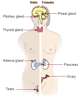
Hormones are responsible for maintaining regular bodily functions, such as growth, digestion, energy, and so on. The endocrine system and the nervous system work together to maintain homeostasis, which is the stable, balanced, and optimal function of the body’s physiological systems. The hypothalamus in the brain regulates our basic biological needs and sends signals to adjust the endocrine system in response to changing needs via the pituitary gland, the “master controller” of the endocrine system, that signals commands to change the production of specific hormones to the other glands in the endocrine system. The table below summarizes the functions of some of the major hormones.
| Hormone | Description and Function | Notes |
|---|---|---|
| Androgens | Masculinizing hormones that cause male sex characteristics; important in sexual drive in both sexes; linked to social aggression and dominance; produced mainly in testes in men, in ovaries in women, and also in adrenal glands. | Testosterone is the most important androgen. |
| Estrogens | Feminizing hormones that cause female secondary sex characteristics to develop at puberty; regulate female fertility linked to learning and memory in both sexes; produced mainly in ovaries in women, in testes in men, and also in adrenal glands. | |
| Adrenal hormones | Help to mobilize the body’s resources; produced by adrenal glands above the kidneys in response to physical or emotional stress or threat; release of cortisol, epinephrine/adrenaline, and norepinephrine is activated by the sympathetic nervous system. | Active in the flight-or-fight response. |
| Endorphins | Reduces pain and induces pleasure; released when under stress; similar effects to natural opiates such as morphine. | Sometimes classified as neurotransmitters or neuromodulators; referred to here as hormones for convenience. |
| Melatonin | Promotes sleep and helps to regulate circadian rhythm. | Released by the pineal gland. |
Key Takeaways
- The central nervous system is the collection of neurons that make up the brain and the spinal cord.
- The peripheral nervous system is the collection of neurons that link the central nervous system to our skin, muscles, and glands.
- Neurons are specialized cells, found in the nervous system, which transmit information. Neurons contain a dendrite, a soma, and an axon.
- Some axons are covered with a fatty substance known as the myelin sheath, which surrounds the axon, acting as an insulator and allowing faster transmission of the electrical signal.
- The dendrite is a treelike extension that receives information from other neurons and transmits electrical stimulation to the soma.
- The axon is an elongated fibre that transfers information from the soma to the terminal buttons.
- Neurotransmitters relay information chemically from the terminal buttons and across the synapses to the receiving dendrites using a system similar to a lock and key.
- The many different neurotransmitters work together to influence cognition, memory, and behaviour.
- Agonists are drugs that mimic the actions of neurotransmitters, whereas antagonists are drugs that block the actions of neurotransmitters.
- Hormones are another type of chemical messenger in the body.
Exercises and Critical Thinking
- Draw a picture of a neuron and label its main parts.
- Test yourself to see if you can identify what is happening in the following YouTube link showing a model of the electrochemical action of the neuron and neurotransmitters:
- Video: Nerve Impulse Animation (Hausmann, 2010)
Image Attributions
Figure 3.1. Used under a CC BY-NC-SA 4.0 license.
Figure 3.2. Used under a CC BY-NC-SA 4.0 license.
Figure 3.3. Used under a CC BY-NC-SA 4.0 license.
Figure 3.4. Major Endocrine Glands by the U.S. Government is in the public domain.
References
CrashCourse. (2015, March 2). The nervous system, part 2 – Action! Potential!: Crash Course A&P #9 [Video file]. Retrieved from https://www.youtube.com/watch?time_continue=72&v=OZG8M_ldA1M
Hausmann, D. (2010, February 14). Nerve impulse animation [Video file]. Retrieved from https://www.youtube.com/watch?v=dSkxlpNs3tU
Khan Academy. (2010, February 11). Anatomy of a neuron | Human anatomy and physiology | Health & medicine | Khan Academy [Video file]. Retrieved from https://www.youtube.com/watch?v=ob5U8zPbAX4
